what would happen if the sister chromatids fail to separate
Affiliate 7: Introduction to the Cellular Basis of Inheritance
7.3 Errors in Meiosis
Learning Objectives
By the cease of this section, you will be able to:
- Explicate how nondisjunction leads to disorders in chromosome number
- Describe how errors in chromosome structure occur through inversions and translocations
Inherited disorders can arise when chromosomes behave abnormally during meiosis. Chromosome disorders tin can exist divided into 2 categories: abnormalities in chromosome number and chromosome structural rearrangements. Because even small-scale segments of chromosomes can span many genes, chromosomal disorders are characteristically dramatic and often fatal.
Disorders in Chromosome Number
The isolation and microscopic observation of chromosomes forms the basis of cytogenetics and is the primary method by which clinicians detect chromosomal abnormalities in humans. A karyotype is the number and appearance of chromosomes, including their length, banding pattern, and centromere position. To obtain a view of an individual'due south karyotype, cytologists photograph the chromosomes and then cutting and paste each chromosome into a chart, or karyogram (Figure 7.7).
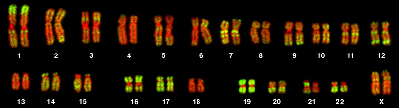
Geneticists Apply Karyograms to Identify Chromosomal Aberrations
The karyotype is a method past which traits characterized by chromosomal abnormalities can be identified from a single cell. To discover an individual'southward karyotype, a person'south cells (like white claret cells) are first collected from a claret sample or other tissue. In the laboratory, the isolated cells are stimulated to brainstorm actively dividing. A chemical is and then applied to the cells to arrest mitosis during metaphase. The cells are and so fixed to a slide.
The geneticist then stains chromosomes with ane of several dyes to better visualize the distinct and reproducible banding patterns of each chromosome pair. Post-obit staining, chromosomes are viewed using bright-field microscopy. An experienced cytogeneticist can identify each ring. In addition to the banding patterns, chromosomes are further identified on the footing of size and centromere location. To obtain the archetype depiction of the karyotype in which homologous pairs of chromosomes are aligned in numerical order from longest to shortest, the geneticist obtains a digital image, identifies each chromosome, and manually arranges the chromosomes into this pattern.
At its most bones, the karyogram may reveal genetic abnormalities in which an individual has too many or too few chromosomes per jail cell. Examples of this are Down's syndrome, which is identified by a third re-create of chromosome 21, and Turner syndrome, which is characterized by the presence of only one 10 chromosome in women instead of two. Geneticists can also identify large deletions or insertions of Deoxyribonucleic acid. For instance, Jacobsen syndrome, which involves distinctive facial features equally well as heart and haemorrhage defects, is identified by a deletion on chromosome 11. Finally, the karyotype can pinpoint translocations, which occur when a segment of genetic material breaks from one chromosome and reattaches to another chromosome or to a different part of the same chromosome. Translocations are implicated in certain cancers, including chronic myelogenous leukemia.
By observing a karyogram, geneticists tin can really visualize the chromosomal limerick of an individual to confirm or predict genetic abnormalities in offspring even before nascence.
Nondisjunctions, Duplications, and Deletions
Of all the chromosomal disorders, abnormalities in chromosome number are the most easily identifiable from a karyogram. Disorders of chromosome number include the duplication or loss of entire chromosomes, as well as changes in the number of complete sets of chromosomes. They are acquired by nondisjunction, which occurs when pairs of homologous chromosomes or sister chromatids fail to separate during meiosis. The risk of nondisjunction increases with the age of the parents.
Nondisjunction tin occur during either meiosis I or II, with different results (Figure 7.8). If homologous chromosomes fail to separate during meiosis I, the result is two gametes that lack that chromosome and two gametes with two copies of the chromosome. If sister chromatids fail to separate during meiosis Two, the effect is ane gamete that lacks that chromosome, two normal gametes with 1 copy of the chromosome, and ane gamete with two copies of the chromosome.
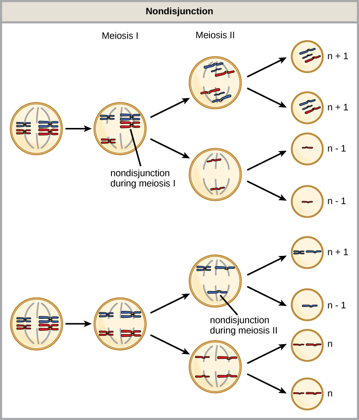
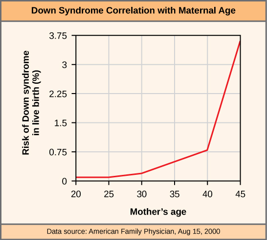
An individual with the advisable number of chromosomes for their species is chosen euploid; in humans, euploidy corresponds to 22 pairs of autosomes and one pair of sex chromosomes. An individual with an error in chromosome number is described as aneuploid, a term that includes monosomy (loss of one chromosome) or trisomy (gain of an inapplicable chromosome). Monosomic human zygotes missing whatsoever one copy of an autosome invariably fail to develop to birth considering they have only 1 copy of essential genes. Most autosomal trisomies likewise fail to develop to birth; withal, duplications of some of the smaller chromosomes (13, fifteen, xviii, 21, or 22) tin can upshot in offspring that survive for several weeks to many years. Trisomic individuals suffer from a unlike type of genetic imbalance: an backlog in factor dose. Cell functions are calibrated to the amount of gene product produced by ii copies (doses) of each cistron; adding a third copy (dose) disrupts this balance. The most common trisomy is that of chromosome 21, which leads to Down's syndrome. Individuals with this inherited disorder have characteristic physical features and developmental delays in growth and cognition. The incidence of Down syndrome is correlated with maternal age, such that older women are more likely to requite birth to children with Downwards syndrome (Figure 7.9).
Concept in Activity
Visualize the add-on of a chromosome that leads to Down syndrome in this video simulation.
Humans brandish dramatic deleterious effects with autosomal trisomies and monosomies. Therefore, information technology may seem counterintuitive that human females and males can function normally, despite carrying unlike numbers of the X chromosome. In office, this occurs because of a process called X inactivation. Early in development, when female person mammalian embryos consist of just a few thousand cells, one 10 chromosome in each cell inactivates by condensing into a structure called a Barr torso. The genes on the inactive X chromosome are not expressed. The particular X chromosome (maternally or paternally derived) that is inactivated in each cell is random, but one time the inactivation occurs, all cells descended from that cell will have the same inactive X chromosome. By this process, females compensate for their double genetic dose of X chromosome.
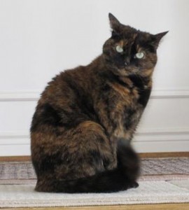
In so-chosen "tortoiseshell" cats, Ten inactivation is observed as coat-color variegation (Figure 7.10). Females heterozygous for an 10-linked coat color cistron will express 1 of two different coat colors over dissimilar regions of their body, corresponding to whichever 10 chromosome is inactivated in the embryonic cell progenitor of that region. When you see a tortoiseshell cat, you will know that information technology has to be a female person.
In an individual carrying an aberrant number of 10 chromosomes, cellular mechanisms volition inactivate all but ane X in each of her cells. Equally a outcome, X-chromosomal abnormalities are typically associated with balmy mental and physical defects, as well as sterility. If the X chromosome is absent birthday, the individual will not develop.
Several errors in sex activity chromosome number have been characterized. Individuals with 3 X chromosomes, called triplo-X, announced female person but limited developmental delays and reduced fertility. The XXY chromosome complement, corresponding to one type of Klinefelter syndrome, corresponds to male individuals with minor testes, enlarged breasts, and reduced body pilus. The actress Ten chromosome undergoes inactivation to compensate for the excess genetic dosage. Turner syndrome, characterized equally an X0 chromosome complement (i.e., just a unmarried sexual practice chromosome), corresponds to a female individual with brusk stature, webbed skin in the neck region, hearing and cardiac impairments, and sterility.
An individual with more than than the right number of chromosome sets (two for diploid species) is called polyploid. For instance, fertilization of an abnormal diploid egg with a normal haploid sperm would yield a triploid zygote. Polyploid animals are extremely rare, with only a few examples amidst the flatworms, crustaceans, amphibians, fish, and lizards. Triploid animals are sterile because meiosis cannot proceed normally with an odd number of chromosome sets. In contrast, polyploidy is very common in the constitute kingdom, and polyploid plants tend to be larger and more robust than euploids of their species.
Chromosome Structural Rearrangements
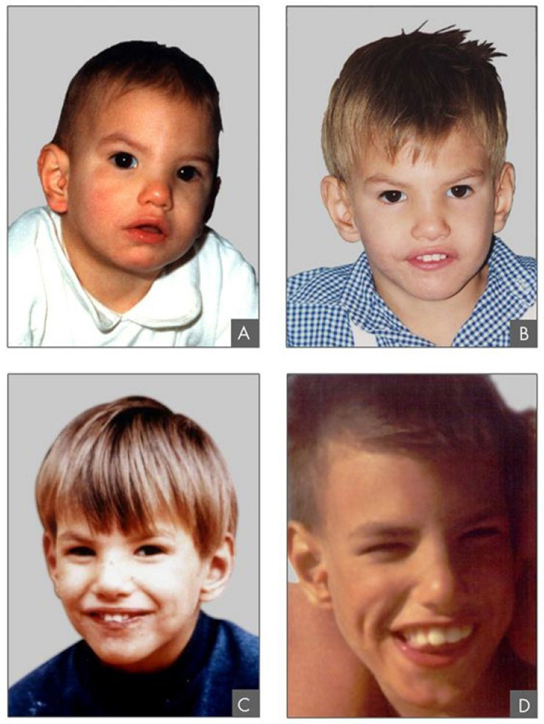
Cytologists have characterized numerous structural rearrangements in chromosomes, including partial duplications, deletions, inversions, and translocations. Duplications and deletions often produce offspring that survive just exhibit physical and mental abnormalities. Cri-du-chat (from the French for "weep of the cat") is a syndrome associated with nervous organisation abnormalities and identifiable physical features that results from a deletion of most of the small arm of chromosome v (Effigy 7.11). Infants with this genotype emit a feature loftier-pitched cry upon which the disorder'due south proper name is based.
Chromosome inversions and translocations can be identified by observing cells during meiosis because homologous chromosomes with a rearrangement in one of the pair must contort to maintain appropriate gene alignment and pair effectively during prophase I.
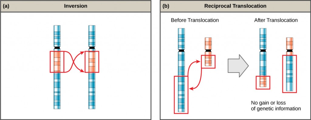
A chromosome inversion is the detachment, 180° rotation, and reinsertion of role of a chromosome. Unless they disrupt a gene sequence, inversions only change the orientation of genes and are likely to have more mild effects than aneuploid errors.
Evolution in Activity
The Chromosome 18 InversionNot all structural rearrangements of chromosomes produce nonviable, dumb, or infertile individuals. In rare instances, such a change can consequence in the evolution of a new species. In fact, an inversion in chromosome 18 appears to take contributed to the evolution of humans. This inversion is non present in our closest genetic relatives, the chimpanzees.
The chromosome 18 inversion is believed to accept occurred in early humans following their divergence from a common ancestor with chimpanzees approximately five million years ago. Researchers have suggested that a long stretch of Deoxyribonucleic acid was duplicated on chromosome 18 of an ancestor to humans, but that during the duplication it was inverted (inserted into the chromosome in reverse orientation.
A comparison of homo and chimpanzee genes in the region of this inversion indicates that two genes—ROCK1 and USP14—are farther autonomously on human being chromosome eighteen than they are on the corresponding chimpanzee chromosome. This suggests that one of the inversion breakpoints occurred betwixt these two genes. Interestingly, humans and chimpanzees express USP14 at distinct levels in specific cell types, including cortical cells and fibroblasts. Perhaps the chromosome eighteen inversion in an ancestral human being repositioned specific genes and reset their expression levels in a useful way. Considering both ROCK1 and USP14 code for enzymes, a change in their expression could alter cellular office. It is not known how this inversion contributed to hominid evolution, just it appears to exist a meaning cistron in the deviation of humans from other primates. 1
A translocation occurs when a segment of a chromosome dissociates and reattaches to a different, nonhomologous chromosome. Translocations can be beneficial or have devastating effects, depending on how the positions of genes are altered with respect to regulatory sequences. Notably, specific translocations have been associated with several cancers and with schizophrenia. Reciprocal translocations result from the exchange of chromosome segments between 2 nonhomologous chromosomes such that there is no gain or loss of genetic information (Figure seven.12).
Section Summary
The number, size, shape, and banding pattern of chromosomes brand them hands identifiable in a karyogram and allow for the assessment of many chromosomal abnormalities. Disorders in chromosome number, or aneuploidies, are typically lethal to the embryo, although a few trisomic genotypes are feasible. Because of X inactivation, aberrations in sexual practice chromosomes typically have milder effects on an private. Aneuploidies also include instances in which segments of a chromosome are duplicated or deleted. Chromosome structures also may be rearranged, for example past inversion or translocation. Both of these aberrations can consequence in negative furnishings on evolution, or death. Because they force chromosomes to assume contorted pairings during meiosis I, inversions and translocations are often associated with reduced fertility because of the likelihood of nondisjunction.
Glossary
aneuploid: an individual with an error in chromosome number; includes deletions and duplications of chromosome segments
autosome: whatever of the not-sex chromosomes
chromosome inversion: the disengagement, 180° rotation, and reinsertion of a chromosome arm
euploid: an private with the appropriate number of chromosomes for their species
karyogram: the photographic image of a karyotype
karyotype: the number and appearance of an individuals chromosomes, including the size, banding patterns, and centromere position
monosomy: an otherwise diploid genotype in which one chromosome is missing
nondisjunction: the failure of synapsed homologs to completely separate and migrate to separate poles during the first prison cell division of meiosis
polyploid: an individual with an incorrect number of chromosome sets
translocation: the procedure past which one segment of a chromosome dissociates and reattaches to a different, nonhomologous chromosome
trisomy: an otherwise diploid genotype in which one entire chromosome is duplicated
X inactivation: the condensation of X chromosomes into Barr bodies during embryonic development in females to compensate for the double genetic dose
Footnotes
1 5 Goidts, et al., "Segmental duplication associated with the human being-specific inversion of chromosome eighteen: a further example of the touch of segmental duplications on karyotype and genome evolution in primates," Man Genetics, 115 (2004):116–22.
Source: https://opentextbc.ca/biology/chapter/7-3-errors-in-meiosis/
Enregistrer un commentaire for "what would happen if the sister chromatids fail to separate"